21+ Tympanic Membrane Color
Transparent Pearl grey in color. A distinctly red tympanic membrane is also helpful adjusted LR 84.

Photographs Retracted Eardrums Retraction Pockets Cholesteatomas Eardrum Perforations Serous And Acute Otitis Media Ear Fluid
A ruptured eardrum tympanic membrane perforation is a hole or tear in the thin tissue that separates the ear canal from the middle ear eardrum.

. Smoking and drinking alcohol increase the risk compared with alcohol use alone. Tympanic membrane eardrum is an oval semi-transparent structure situated between the and the tympanic cavity of the. Use the largest ear speculum that will fit to give maximal visualization and optimal seal 2 and 4 mm sizes are common III.
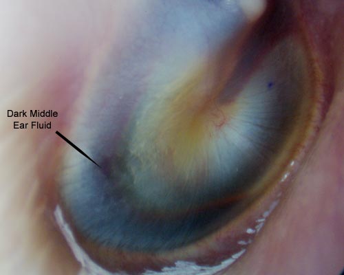
The blue ear drum generally refers to a condition in which blood or blood products are found in the middle ear. A tympanic membrane that has normal color and mobility is not typical for acute otitis media. After all possible causes for hemotympanum including blood dyscrasias and.
Chronic mastoiditis is defined by the presence of long-standing infection in the presence of. Diagnosis Treatment Myringosclerosis and tympanosclerosis are similar conditions that affect the middle ear causing the tympanic membrane eardrum to appear bright white. Detecting the Likelihood of Acute Otitis Media Healthy children who cry before and during the examination are unlikely to have distinctly red tympanic membranes.
Although 80 of cases resolve spontaneously in the US antibiotics are often given 1 Treatment. At a distance of 14-16 inches refraction is Ability of the eye to bend light rays so that they can be focused on the retina After performing an ear instillation the patient is instructed to lie on the. A ruptured eardrum can.
Tilted at an angle of to the ear canal. When viewed through the overlying tympanic membrane this blood usually appears purple blue brown or gray. Topical agents should not be used when there is a tympanic membrane perforation.
At the Tympanic Membrane TM hence defines the border between the outer and middle ear. A rare consideration is a middle ear mass such as a meningocele or. Read chapter 23 of The Color Atlas of Pediatrics online now exclusively on AccessPediatrics.
95 CI 67-11 whereas a normal color makes AOM much less likely adjusted LR 02. The tympanic membrane is divided into.

Heent Conditions Flashcards Quizlet

Photographs Retracted Eardrums Retraction Pockets Cholesteatomas Eardrum Perforations Serous And Acute Otitis Media Ear Fluid

Inspect The Tympanic Membrane Physical Diagnosis Mitch Medical
Ear Trauma Images Mcgovern Medical School
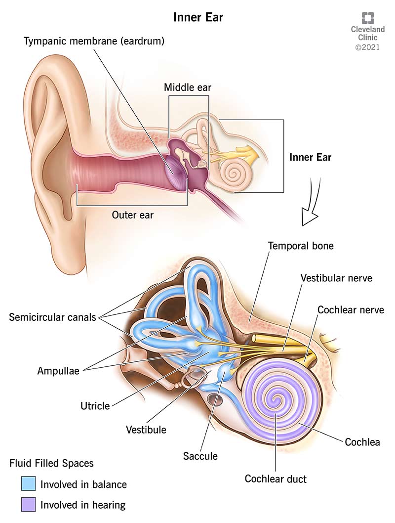
Inner Ear Anatomy Function

Eardrum Color And The Imaging Diagnosis Of Middle Ear Disease Otoscopic Radiologic Correlation Of Retrotympanic Lesions Semantic Scholar

Otoscopy Pathologies

Color Atlas Of The Anatomy And Pathology Of The Epitympanum Karger Publishers
Tympanic Membrane Pictures Function Anatomy Body Maps

Automated Diagnosis Of Ear Disease Using Ensemble Deep Learning With A Big Otoendoscopy Image Database Ebiomedicine

Illustrated Intro To Middle Ear Anatomy As Seen By Otoscopy Wiscmed

Tympanic Membrane Pictures Function Anatomy Body Maps

Eardrum Color And The Imaging Diagnosis Of Middle Ear Disease Otoscopic Radiologic Correlation Of Retrotympanic Lesions Semantic Scholar

Left Tympanic Membrane With Features Of A Normal Middle Ear The Handle Download Scientific Diagram

Eardrum Color And The Imaging Diagnosis Of Middle Ear Disease Otoscopic Radiologic Correlation Of Retrotympanic Lesions Semantic Scholar

Normal Tympanic Membrane The Normal Tympanic Membrane Appears As A Pale Download Scientific Diagram

Otoscopy Pathologies